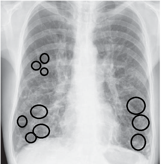

This is caused by irregularly piled-up mucus interspersed with air pockets. On imaging studies like CT scans the bronchi may also appear beaded like a rosary. However, occasionally there may be appearance of thickening of bronchial walls and crowding of bronchial structures in the lower part of the lungs on X ray in severe cases. Physical examination may also show lack of oxygen persistent leading to cyanosis (blue lips and nail beds).Ĭhest X ray may be normal in mild chronic bronchitis. Wheezing may be noted on inspiration or expiration and expiration frequently is prolonged.

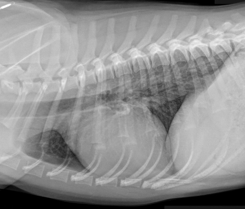
On hearing the breath sounds with a stethoscope, the breath sounds rough or harsh and raspy with coarse sounding rales, rhonchi etc. These patients however may have a history of previous episodes of flare ups and recurrent episodes of acute bronchitis. Physical examination may not reveal any abnormality specifically. Pulse oximetry is recommended to check the blood oxygen levels. These tests are needed in recurrent bronchitis cases. Lung function tests like spirometry are not routinely used in the diagnosis of acute bronchitis. A chest X ray may also be advised to elderly patients, those with chronic obstructive pulmonary disease, recent episode of pneumonia, cancer, tuberculosis patients and those with debilitated status or lowered immunity. This however, is not very conclusive in acute bronchitis since most cases are caused by respiratory viruses.Ī chest X ray is performed in patients whose physical examination suggests pneumonia or co-existing heart failure. Sputum may be tested in laboratories for microbes. High persistent fever however indicates pneumonia or influenza. The physical examination of patients presenting with symptoms of acute bronchitis reveals presence of fever, rapid breathing rate, wheezing, noisy respiration, rapid heart rate, prolonged and noisy expiration etc.įever may be present in some patients with acute bronchitis. Diagnosis is based on clinical history, physical examination as well as laboratory, imaging as well as breath analysis tests. Ananya Mandal, MD Reviewed by April Cashin-Garbutt, MA (Editor)īronchitis is mainly caused by a chest infection that leads to pathological changes in the narrow airways of the lungs.


 0 kommentar(er)
